
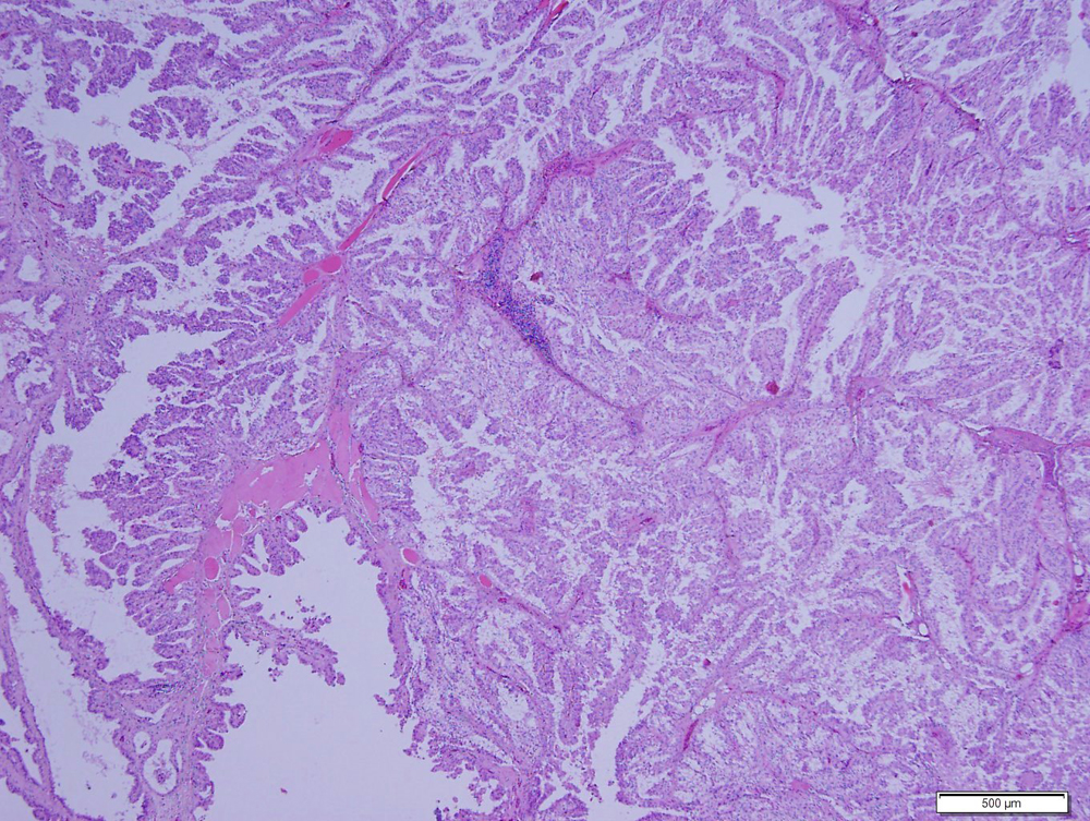
The tumor is comprised of papilliform arrangements of epithelial cells, which are generally one layer thick in this image. The epithelial cells are supporting by a thin fibrovascular stroma, corresponding to the matrix seen in the cytologic smears (H&E stain, 4x objective)