
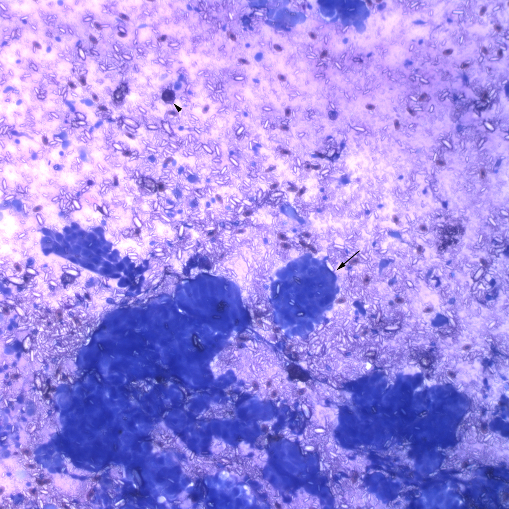
A direct smear of the fluid aspirated from the swelling consisted of numerous clusters (arrow) and a few flat sheets of medium to large epithelial cells with many extracellular crystals and low numbers of macrophages, which were phagocytizing crystals (arrowhead) (modified Wright’s stain, 10x objective).