Acute phase proteins (APPs) are defined as proteins that change their serum concentration by >25% in response to inflammatory cytokines (IL-1, IL-6, TNFα). The acute-phase response is considered part of the innate immune system, and APPs play a role in mediating such systemic effects as fever, leukocytosis, increased cortisol, decreased thyroxine, decreased serum iron, and many others. APPs can be categorized as positive (increasing serum concentration) or negative (decreasing serum concentration).
Increased production of positive acute phase proteins is a sensitive indicator of inflammation which can occur prior to the development of an inflammatory leukogram.
| Positive APPs | Negative APPs |
| C-reactive protein (CRP) | Albumin |
| Serum Amyloid A (SAA) | Transferrin |
| Haptoglobin (Hp) | Transthyretin |
| Ceruloplasmin | Retinol-binding protein |
| α2-Macroglobulin | Adiponectin |
| α1-Acid glycoprotein (AGP) | |
| Fibrinogen | |
| Complement (C3, C4) |
General
Positive acute phase proteins
Positive acute-phase proteins increase in plasma concentration in response to inflammation (usually within 1-2 days). Positive APPs are further categorized as major, moderate or minor, depending on the degree of increase.
- Major APP: A protein with a low concentration in the serum of healthy animals (often <0.1 μg/dL), but upon stimulation will increase over 100 – 1000 fold, reaching a peak 24-48 hours after insult, then rapidly decreasing. An example of an major APP is Serum amyloid A.
- Moderate APP: Present in the blood of healthy animals, but increases 5 – 10 fold upon stimulation, peaking around 48 – 72 hours after insult, then decreases at a slower rate than major APPs.
- Minor APP: Increase only by 50 – 100% above resting levels and at a gradual rate.
The rapidity and magnitude of the increase in each acute phase protein varies depending on the species. The following table list the acute phase proteins that are major and moderate responders in various animal species.
| Species | Major APP | Moderate APP |
| Cat | SAA | AGP, Hp |
| Dog | CRP, SAA | Hp, AGP, Cp |
| Horse | SAA | Hp |
| Cow | Hp, SAA | AGP |
| Pig | CRP, Pig-MAP | Hp, Cp |
| Mouse | SAA | Hp, AGP |
| Rat | α2-macroglobulin | Hp, AGP |
| SAA = Serum amyloid A; CRP = C-reactive protein; Hp = Haptoglobin; Pig-MAP = Major acute phase protein; | ||
Acute-phase proteins are part of the innate immune response and its biological function, although variable, generally relate to defense to pathological damage and restoration of homeostasis. However, a specific APP may have both pro- and anti-inflammatory effects. The following table summarizes the functions of the major APPs.
| Protein | Main function |
| Alpha-1-acid glycoprotein | Antiinflammatory and immunomodulatory agent: has antineutrophil and anticomplement activity and increases macrophage secretion of IL-1 receptor antagonist. Binds to lipophilic and acidic drugs. |
| C-reactive protein | On bacteria, it promotes the binding of complement, facilitating phagocytosis. Induction of cytokines Inhibition of chemotaxis and modulation of neutrophil function Neutralizes deleterious effects of histones |
| Ceruloplasmin | Copper transport (for wound healing, collagen formation and maturation) Antioxidant Reduces the number of neutrophils attaching to endothelium |
| Haptoglobin | Binds free hemoglobin (limiting Hb iron availability for bacterial growth) Natural antagonist for receptor-ligand activation of the immune systemInhibition of granulocyte chemotaxis and phagocytosis |
| Serum amyloid A | Chemotactic recruitment of inflammatory cells to sites of inflammation Induction of inflammatory cytokines (via surface receptors, including Toll like receptor) Inhibition of myeloperoxidase release and lymphocyte proliferation Involved in lipid metabolism and transport immunomodulatory (via the inflammasome) |
Negative acute phase proteins
Negative acute phase proteins decrease in plasma concentration by greater than 25% in response to inflammation. This reduction can occur rapidly (within 24 hours) or may decrease gradually over a period of days. The two main negative acute phase proteins are albumin and transferrin. The mechanism by which their concentrations decrease is likely multifactorial, including decreased production by the liver in response to inflammatory cytokines, and possibly increased loss or increased proteolysis.
- Albumin
- Reduced production of albumin allows greater increase in the amount of amino acids available for positive APP production
- Albumin concentration falls gradually and reduction in concentration is more noticeable in chronic inflammatory disease
- Transferrin
- Usually measured to assess iron status
- Ovotransferrin is the avian analog, but it is a positive acute phase protein
- Adiponectin: This protein, which is produced in adipose tissue, and promotes energy usage through increasing sensitivity to insulin, has anti-inflammatory properties. Concentrations may decrease in blood in dogs with inflammation, particularly that due to sepsis. The mechanism of decrease is unclear and is not specific for sepsis, since decreased concentrations may be seen in obese animals or animals with diabetes mellitus (Torrente et al 2020).
Measurement
Serum electrophoresis
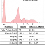
The acute-phase proteins migrate in the α- (mostly) and β- regions of the electrophoretogram. As many acute-phase proteins are α globulins, an increase in concentration of α1 or α2 globulins is a sign of an acute-phase response and is detectable soon after the onset of inflammation, injury, or infection and may persist until the inciting stimulus has resolved.

The typical findings on SPE of the acute-phase response are:
- Normal to mild increase in total protein (included on the SPE report, but measured on the chemistry analyzer)
- Normal to mild decrease in albumin (negative acute-phase protein)
- Variable increase in α1- or α2 globulins
The following table show the electrophoretic region in which specific APPs appear in the electrophoretogram.
| Serum protein | Electrophoretic region |
| α1-Acid glycoprotein | α1 |
| Serum amyloid A | α |
| Haptoglobin | α2 |
| Ceruloplasmin | α2 |
| Transferrin | β1 |
| C-reactive protein | γ |
An an increase in α-globulins may only be observed when acute-phase proteins normally found in high concentrations (milligram or gram quantities, e.g. haptoglobin) are increased in serum. Acute-phase proteins found in smaller amounts (nanogram or picogram quantities, e.g. serum amyloid A) will not result in an increase in α-globulins, even when markedly increased in serum.
Measurement of specific acute-phase proteins is a more sensitive test of the acute-phase response than electrophoresis.
C-reactive protein
- Sample considerations
- Storage: stable at -10 C for 3 months
- Anticoagulant: Do not use citrate tube as levels are significantly lowered
- Tests
- Turbidimetric immunoassay: used in humans and has been adapted for automated biochemical analyzers. However, there is variation in cross-activity with different antihuman CRP antibodies. Hemolysis will interfere with immunoturbidimetric testing.
- ELISA: a commercially available kit for canine CRP
- Slide/capillary reverse passive latex agglutination tests
- Time-resolved fluorometry (TRFIA): recently developed for CRP assays in canine whole blood, saliva and effusions
Ceruloplasmin
- Sample considerations
- Anticoagulant: concentrations are higher with heparin and lower with EDTA
- Tests
- There are problems with ceruloplasmin assays due to the lack of commercially available reference materials to standardize ceruloplasmin concentrations. Therefore, different arbitrary units based on increased absorbance per unit time have been used (oxidase units UI/L).
Haptoglobin
- Sample considerations
- Storage: more stable in serum than in a purified preparation. Values decrease in serum stored at -20 C. Storage at -70°C is recommended.
- Anticoagulants: concentration is increased with heparin
- Interferences: Haptoglobin levels are induced by steroids, so dogs with high endogenous steroids or on exogenous steroid therapy will have increase Hp concentrations. Canine serum specimens must be diluted when Hp assays developed for other species are used. This is because canine Hp concentrations in health or disease are significantly higher than other species.
- Tests
- Spectrophotometric assays
- Hemoglobin-haptoglobin complexes that alter the absorbance characteristic of Hb in proprotion of the concentration of Hb in serum
- Preserve peroxidase acitivity at an acidic pH, which can then be detected and quantified.
- Multispecies assay based on the peroxidase activity of Hb-Hp complexed (interference by albumin is eliminated) has been validated for canine serum
- Immunoassays
- Nephelometric assay: rate of precipitation of the antigen-antibody comples is measured. This has been validated for estimation of haptoglobin in dogs.
- Spectrophotometric assays
Alpha-1 acid glycoprotein
- Tests:
- Estimation by precipitation of majority of serum proteins by perchloric acid and quantification of the remaining soluble proteins.
- Single radial immunodiffusion on agarose gel impregnated with anti-species AGP rabbit serum. Dog and cat specific assays have been developed.
- Immunoturbidimetric assays have been developed for canine and feline AGP measurement
