
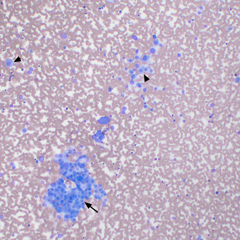
Another low power image showing flat sheets of cells, which appear cohesive (arrow) and individualized more round cells (arrowheads) with bare nuclei (modified Wright stain, 10x objective). This cytologic pattern is typical of endocrine or neuroendocrine tumors.