
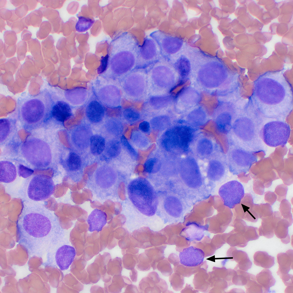
This grouping of neoplastic cells appears more cohesive and the cells have distinct cell boundaries. A few bare nuclei are seen around the edges (arrows) (modified Wright stain, 50x objective).
This grouping of neoplastic cells appears more cohesive and the cells have distinct cell boundaries. A few bare nuclei are seen around the edges (arrows) (modified Wright stain, 50x objective).
eClinpath helped 1.2 million visitors last year from 220 countries find important information on animal health. If you enjoy the site, please support our mission and consider a small gift to help us keep pace with its rapid growth. You can donate securely via PayPal or credit card. Thank you!
![]()