
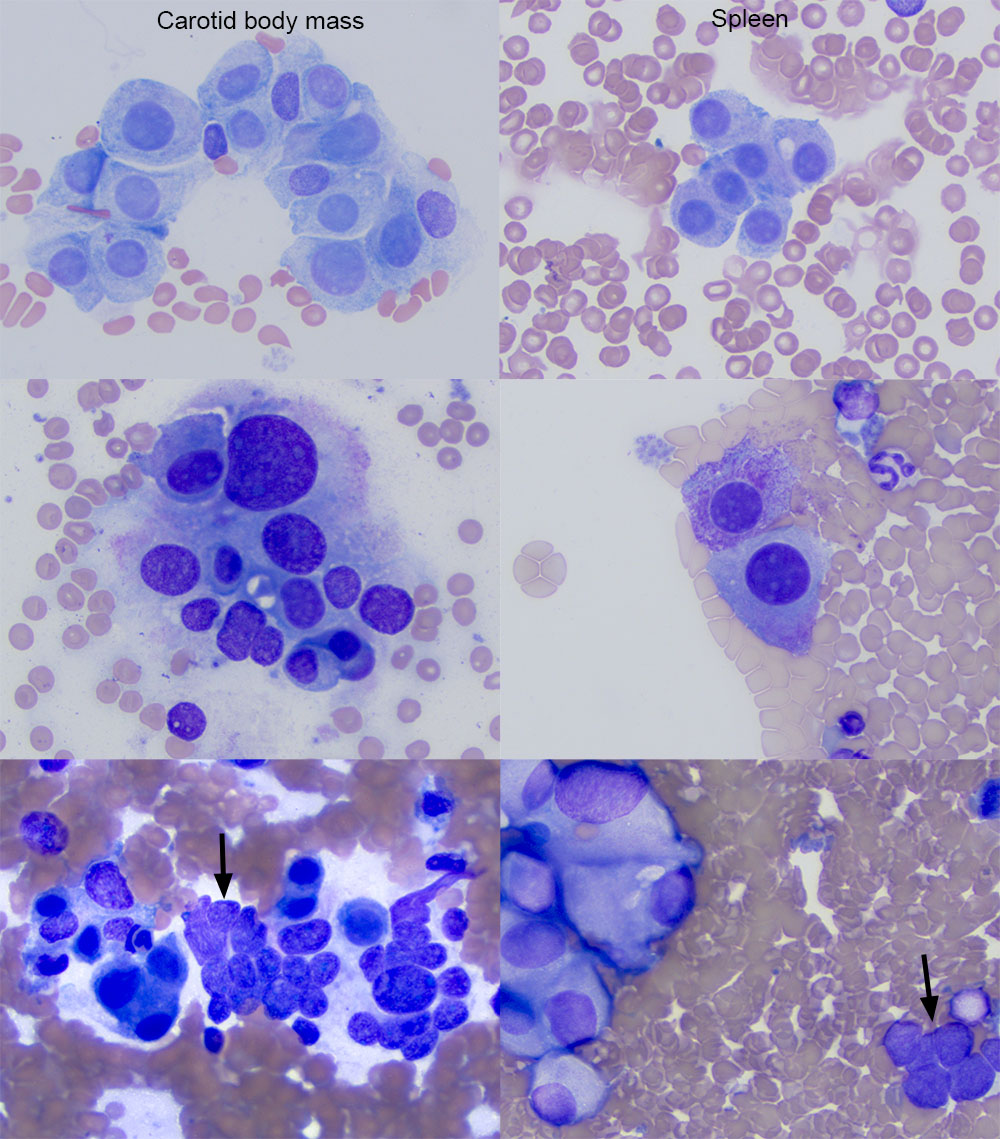
The cells in smears from the aspirate of the mass in the region of the carotid body closely resemble those seen in the aspirate smears of the spleen, including pink-red cytoplasmic granules (second row) and ruptured cells with bare nuclei in groupings (arrows, third row) (modified Wright stain).