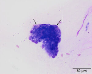
On the edge of a cluster of cuboidal epithelial cells with light to medium blue cytoplasm, there are two cells with eccentric nuclei and numerous pink cytoplasmic granules (arrows). The cytologic features of the cells and their location alongside cuboidal epithelial cells are compatible with parietal cells, which secrete hydrochloric acid in the stomach. These cells are further support for a stomach origin for the epithelial cells in the aspirate from the mid-esophageal nodule Modified Wright’s stain (50x objective)

