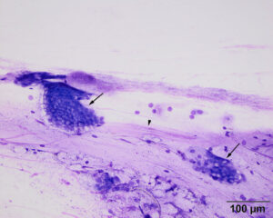
The direct smears contained clusters of mucinous columnar epithelial cells (arrows) embedded in thick strands of mucus (arrowhead). A higher power image of the cells is shown in Figure 3. These cells are an abnormal finding for this region of the esophagus, which should consist of non-keratinized stratified squamous epithelium. The purple granules in the background could be mucin but also resemble the granules seen in chief cells (see Figure 5). Modified Wright’s stain (20x objective)

