
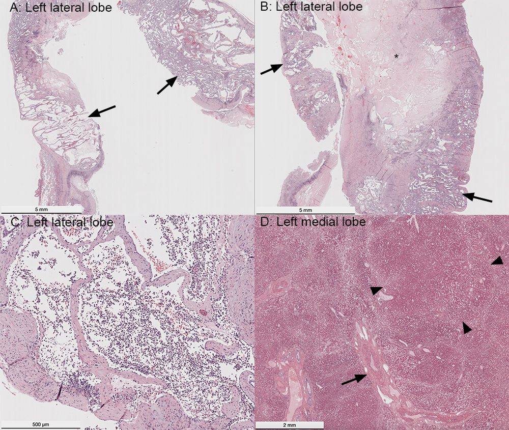
A: A section of the large cystic mass in the left lateral liver lobe revealed numerous dilated and torturous trabeculae of cuboidal epithelial cells, compatible with biliary epithelium (arrows). The center of the mass consisted of a large cyst, which contained neutrophils, sloughed epithelial cells, cellular debris, and erythrocytes in the center. B: Another section of the left lateral liver lobe showed a large area of necrosis (*) and anastomosing and variably dilated trabeculae and cords of epithelial cells (arrows). C: Another area of the mass consisted of cystic dilated regions of epithelial cells supported by a thick fibrous stroma. Note the marked neutrophilic inflammation in the center of the cystic regions. The histologic diagnosis for the mass was a biliary hamartoma (von Meyenburg complex). D: A section of the left medial liver lobe showed irregular nodular formation (arrowheads) and prominent peribiliary fibrosis (arrow). Hematoxylin-&-eosin stain.