
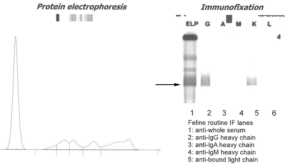
Left panel: Repeat serum electrophoresis done at Colorado Sate University shows a faint monoclonal peak in the far gamma region. The electrophoresis was done using an agarose gel technique, which does not delineate peaks as clearly as capillary zone electrophoresis used at our institution (see Figure 6). Right panel: Gel of the immunofixation performed at Colorado State University, showing immunostaining with antibodies against serum (lane 1), heavy chain of all three types of immunoglobulins (IgG, A and M heavy chain, lanes 2-4) and light chain bound to heavy chain as part of intact immunoglobulins (lane 5). A narrow peak in the gamma and bound light chain lanes (arrow) supports a monoclonal gammopathy. Note the broad smearing of IgG in lane 2, compatible with the polyclonal base identified on electrophoresis. Neither IgA or IgM were present, which could reflect concurrent immunosuppression.