
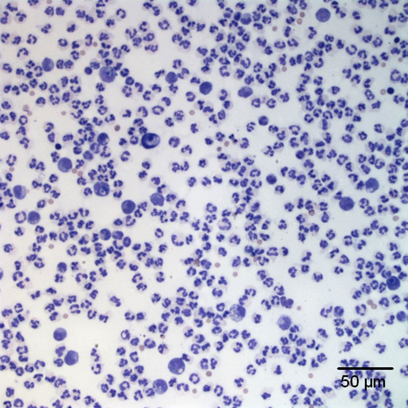
Figure 1a. Exudative effusion. Highly cellular sample consisting of mixed inflammatory cells (>80% neutrophils) on a background of a small amount of blood. (Wright’s stain, 20x objective).
Figure 1a. Exudative effusion. Highly cellular sample consisting of mixed inflammatory cells (>80% neutrophils) on a background of a small amount of blood. (Wright’s stain, 20x objective).
eClinpath helped 1.2 million visitors last year from 220 countries find important information on animal health. If you enjoy the site, please support our mission and consider a small gift to help us keep pace with its rapid growth. You can donate securely via PayPal or credit card. Thank you!
![]()