
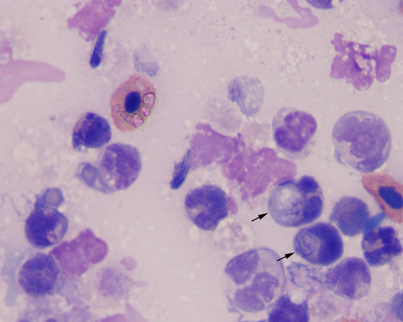
Figure 3: Numerous Spironucleus trophozoites are present amidst degenerating leukocytes and cellular debris in this smear of the fluid from the cystic lesion. Phagocytized organisms were easily found (arrows) (1000x, Wright’s stain).
Figure 3: Numerous Spironucleus trophozoites are present amidst degenerating leukocytes and cellular debris in this smear of the fluid from the cystic lesion. Phagocytized organisms were easily found (arrows) (1000x, Wright’s stain).
eClinpath helped 1.2 million visitors last year from 220 countries find important information on animal health. If you enjoy the site, please support our mission and consider a small gift to help us keep pace with its rapid growth. You can donate securely via PayPal or credit card. Thank you!
![]()