
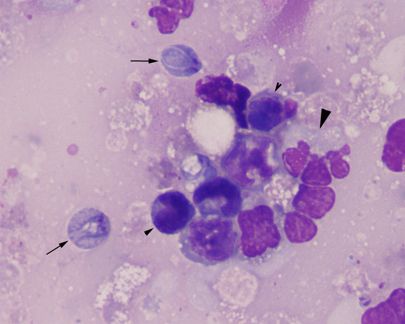
Figure 2: In this higher magnification image, the protozoal trophozoites (arrows) are observed alongside inflammatory cells, including granulocytes (large arrowhead) and lymphocytes (small arrowhead). Flagella are barely discernible against the proteinaceous background (1000x, Wright’s stain).