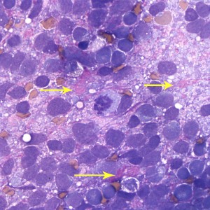
Figure 2a. Neoplastic cells are embedded within a fibrillar to granular to globular bright pink extracellular matrix (arrows). A mitotic figure is evident in the center and a few plasma cells and vacuolated microglia are seen between the tumor cells (Wright’s stain, 500x).

