
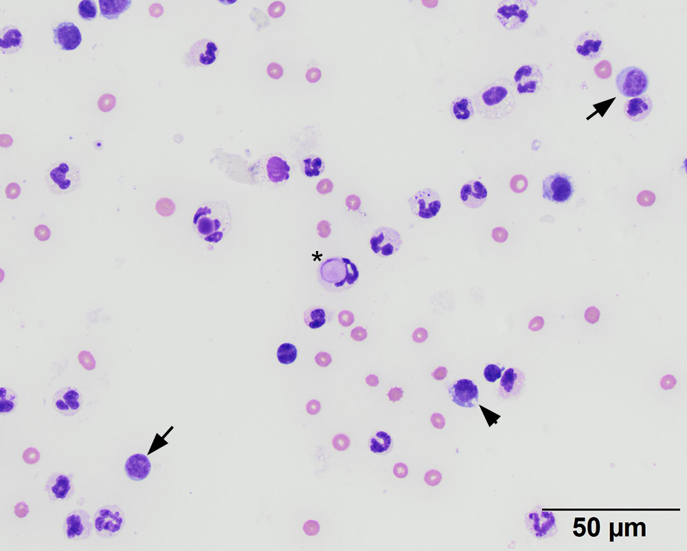
The sediment smear was highly cellular, composed predominantly of neutrophils with fewer lymphocytes (arrows, also called “small mononuclear” cells) and macrophages or synovial lining cells (arrowhead, also called “large mononuclear” cells). A neutrophil containing a light purple large round cytoplasmic inclusion is consistent with a “lupus erythematosis” cell (*). There was a small amount of blood. Cells are not windrowing (lining up), consistent with the visual estimate of reduced viscosity (50x objective, modified Wright stain)