
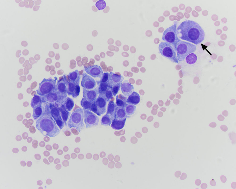
This higher power view shows that the cells aspirated from the mass in the carotid region are similar to those in the smears of the splenic aspirate, confirming this site as the primary tumor. Note the fine pink-red granules in one of the cells (arrow). The cytologic diagnosis was a chemodectoma (at the time of this cytologic diagnosis, the clinical pathologist was unaware it was the same case as the metastatic splenic tumor) (modified Wright stain, 50x objective).