
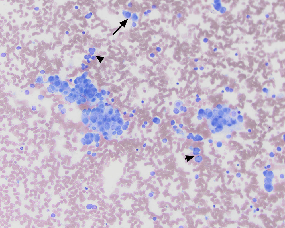
Direct smears of the mass contained packets and clusters of round to oval cells, resembling those seen in the splenic aspirate. Note the individualized cells (arrows) and bare nuclei (arrowheads), some of which are in groupings (modified Wright stain, 20x objective).