
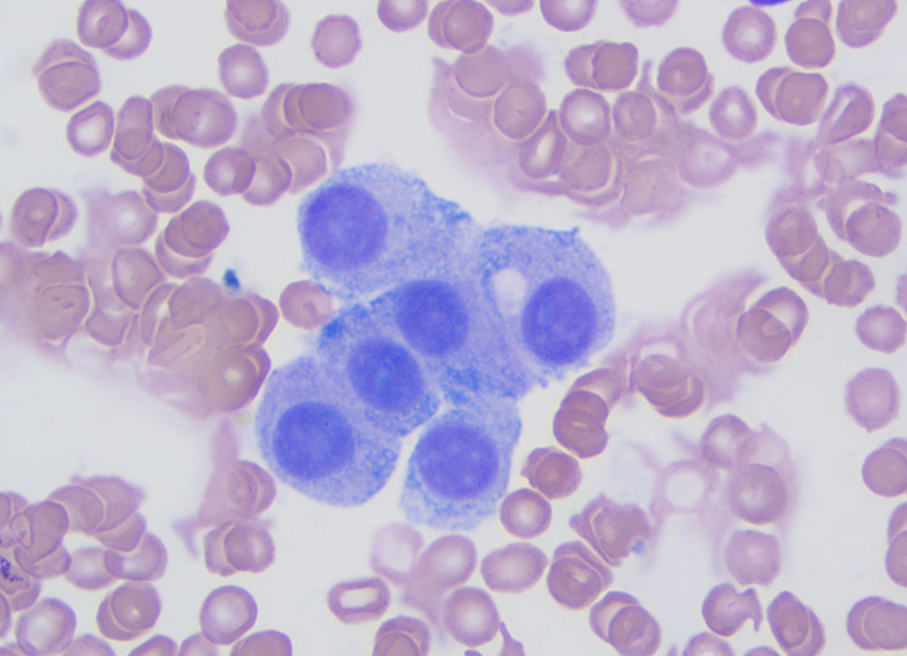
A small cluster of cells, with distinct cell boundaries, and fine blue-green cytoplasmic granules at the periphery of the cells. We have seen these granules in various types of neuroendocrine and endocrine tumors and it is a helpful feature confirming tumor type, but not origin. The red granules shown in image 2c are far less common in these tumors (modified Wright stain, 100x objective).