
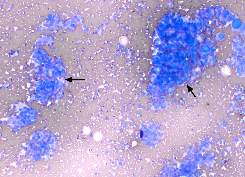
The aspirate of spleen contained multifocal aggregates of cells with intercalated red blood cells, features that we typically associate with mesenchymal tumors (arrows). The cells show mild to moderate anisocytosis and anisokaryosis overall, however you can see individualized cells with macronuclei (modified Wright stain, 20x objective).