
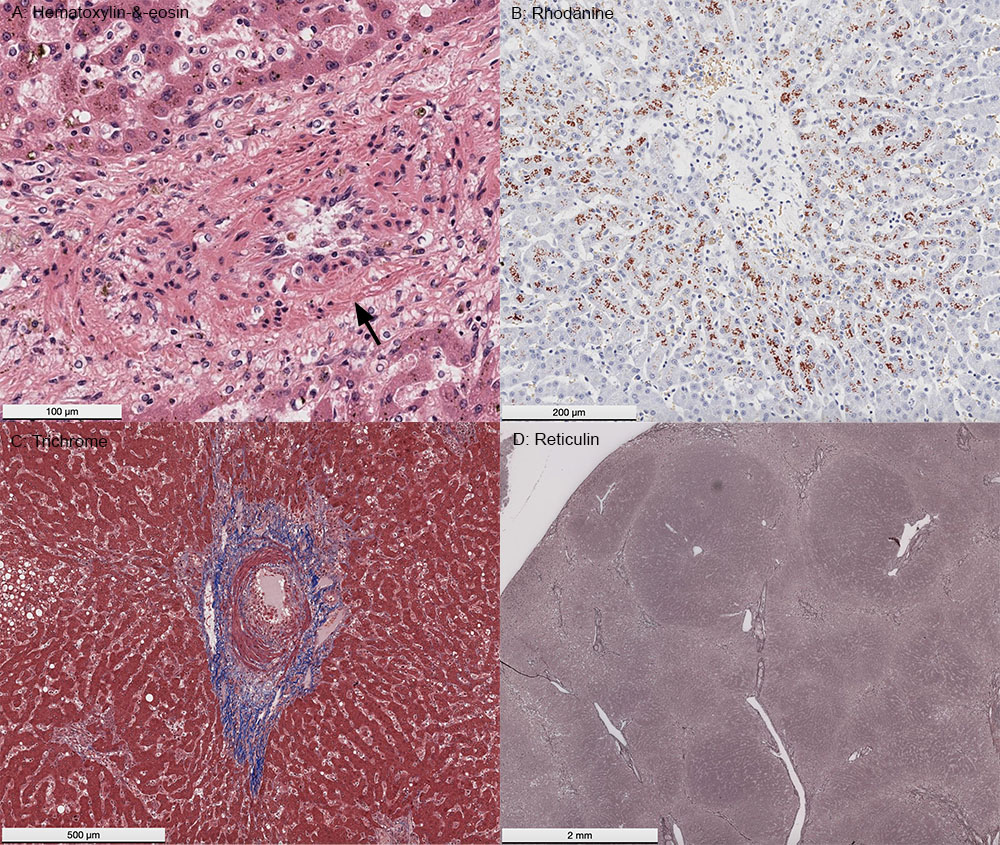
A: Hematoxylin-&-eosin stain of the left medial liver lobe: A higher power image showing the prominent peribiliary fibrosis (arrow) and red-brown pigment in periportal hepatocytes, compatible with copper. B: Rhodanine stain for copper: The pigment in hepatocytes stained with rhodanine, confirming that it was copper. C: Trichrome stain for collagen: The trichrome stain highlights the peribiliary fibrosis. D: Reticulin stain for type III collagen: The reticulin stain highlights the indistinct nodule formation, which likely explained the variably echogenic nodules described on ultrasonographic examination of the liver lobes.