
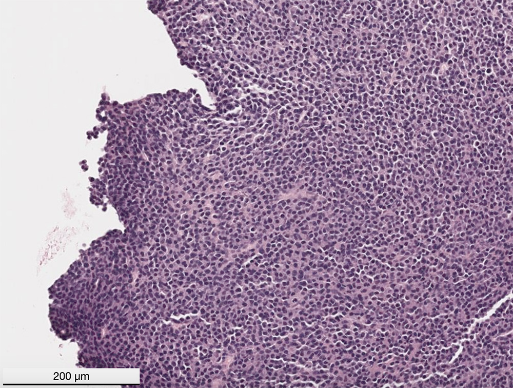
This captured image of the digitized slide of a cryostat section of the mass revealed sheets of round cells supported by fine stroma. The cells extended to the cut margins and infiltrated muscle and bone marrow (not shown). The cells had eccentric nuclei with stippled to clumped chromatin and no nucleoli. They had a moderate amount of amphophilic cytoplasm with a perinuclear clear zone, which are features compatible with plasma cells (H&E stain).