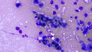
Scraping prepared from a biopsy of a liver mass in a horse: There are several small flat sheets of larger polygonal hepatocytes with central nuclei and pink cytoplasmic granulation. The hepatocytes contain moderate amounts of green-brown to green pigment in their cytoplasm (bilirubin or lipofuscin, presumptive) and bile casts are present (arrow) between some cells, indicating cholestasis (50x objective, Wright’s stain).

