
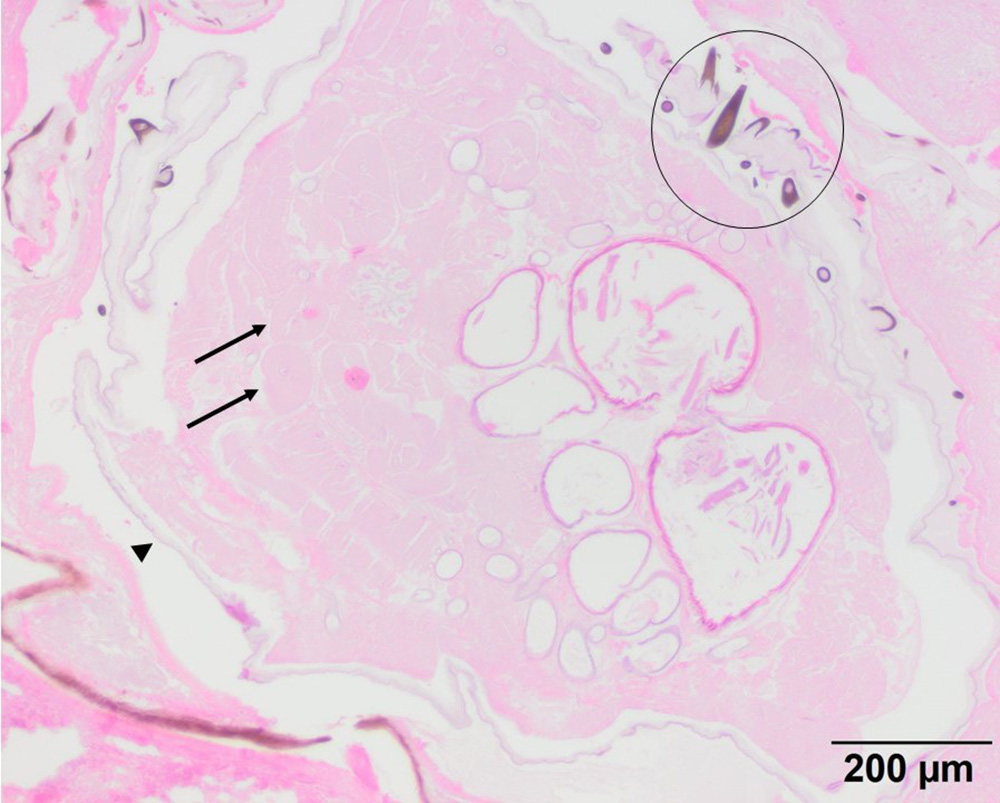
The larva is surrounded by a thin cuticle (black arrowhead) with attached single-pointed brown spines (black circle). The latter are consistent with larvae of the rodent bot fly (Cuterebra). Internal tissue cells are similar to those seen in the cytologic smear (long black arrows) (H&E stain, 10x objective). This case is more than 20 years old, so the histologic section has faded and nuclei in the cells are difficult to discern. The cells are easier to picture in the original publication, albeit in black and white and are of unclear embryonal origin), but could be muscle being associated with the body wall of the larva.