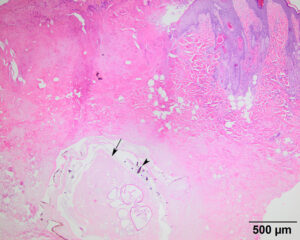
Tissue section from the excisional surgical biopsy of the left mandibular mass. The mass consisted of necrosis with neutrophilic inflammation surrounding a central structure, consistent with an arthropod larva (arrow). Note the melanin-containing single-pointed brown spines on the cuticle of the larva (arrowhead). The surface epithelium was ulcerated , likely associated with central respiratory pore formation (H&E stain, 4x objective).

