
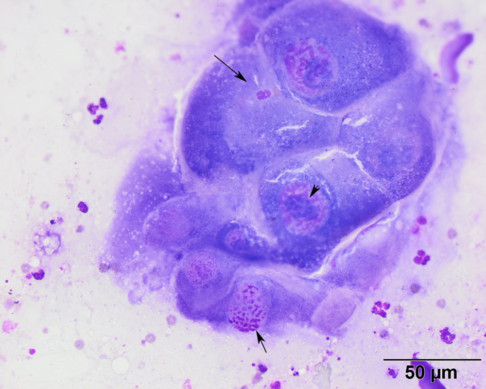
This higher power view of the polygonal cells shows their large size and prominent nucleoli (arrowhead) with ropey chromatin (short arrow). Neutrophil emperipolesis can be seen in one of the cells (long arrow), indicating that the cells were a source of inflammation (modified Wright’s stain, 50x objective)