
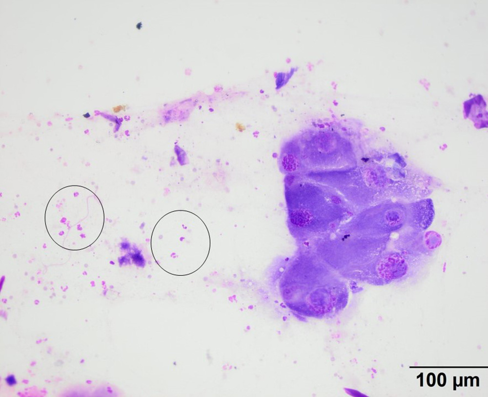
The direct smear of the aspirate consisted of very large polygonal cells (75-100 um), found in clusters, with a few individualized cells, along with moderate numbers of neutrophils, which were mildly to not degenerate (circles). The cells had eccentric large nuclei with ropey to stippled chromatin with single large nucleoli. They had a moderate to large amount of smooth to grainy mid-blue cytoplasm (modified Wright’s stain).