
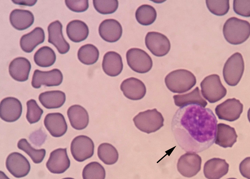
A large oval smooth light pink inclusion is seen within the cytoplasm of the lymphocyte (arrow). Surrounding erythrocytes have some colorless inclusions. These may be artifacts versus virus inclusions. A single large platelet is also present in this smear (Diff-quik, high magnification).