
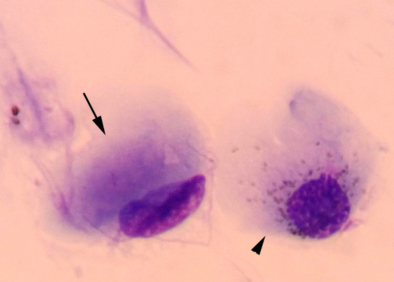
One of the conjunctival epithelial cells appears to contain a large cytoplasmic inclusion with slight pink granularity (arrow). The adjacent epithelial cells contain numerous small brown cytoplasmic granules, compatible with melanin (arrowhead), indicating areas of pigmentation within the conjunctiva, which is likely normal and not related to the canine distemper virus infection (Diff-quik).