
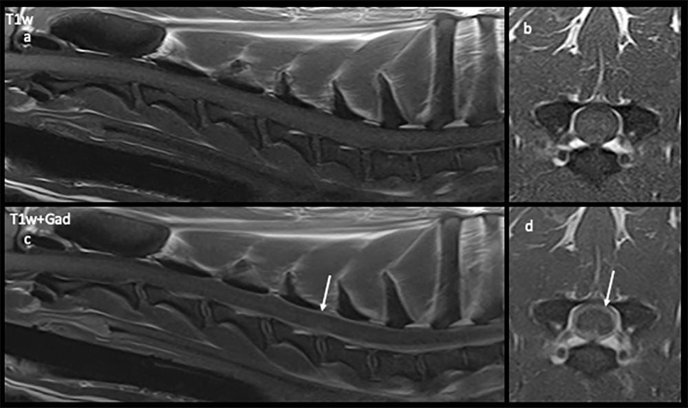
T1-weighted saggital (a: non-contrast; c: contrast) and transverse (b: non-contrast; d: contrast) magnetic resonance images taken at the level of the 5th cervical vertebra. Contrast uptake and hyperintensity in the meninges is evident in the images post injection of contrast (arrows) versus the non-contrast images.