
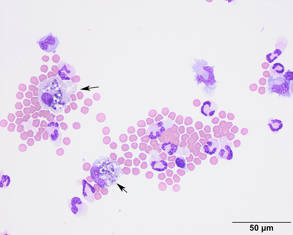
Another image of the cytospin smear from the LS tap shows a phagocytic macrophage with pigmented cytoplasmic globules, considered to be a mixture of phagocytic debris and hemosiderin (arrows). There are also two lymphocytes, several non-degenerate neutrophils, non-phagocytic/non-vacuolated macrophages and red blood cells in this image (modified Wright’s stain).