
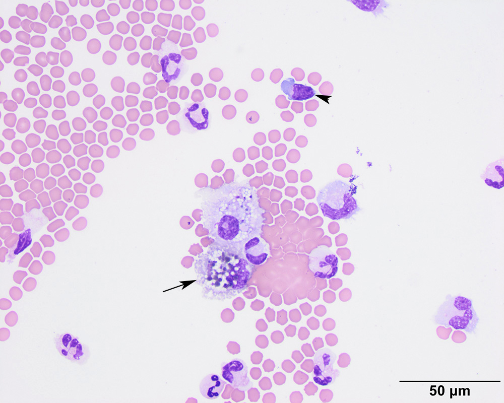
Compared to the smear from the atlanto-occipital tap, there are numerous red blood cells in the background. In addition, macrophages demonstrate phagocytic activity (arrows). Some of the pigmented globules were thought to be compatible with hemosiderin, supporting hemorrhage. A small lymphocyte is reactive, with bluer cytoplasm than normal (arrowhead) (modified Wright’s stain).