
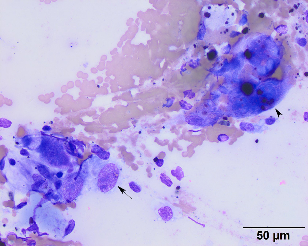
Two osteoclast-like giant cells, containing erythrocytes and hemosiderin in their cytoplasm, are seen in this image (one labeled with an arrowhead), along with spindle cells showing cytologic criteria of malignancy (large oval nucleus with two nucleoli of irregular shape and size, arrow) (50x objective, modified Wright’s stain).