
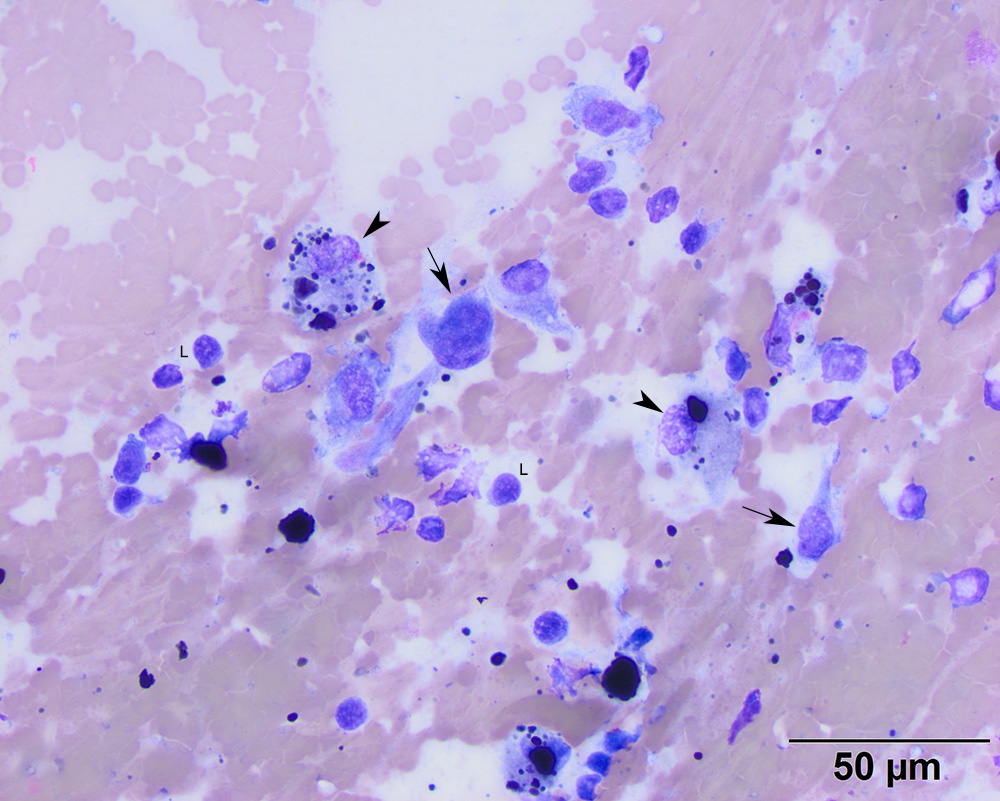
A higher power view of the smear shows that small lymphocytes (L) dominate, with moderate to many macrophages, which were erythrophagocytic (not shown) and/or contained hemosiderin in their cytoplasm (arrowheads). There was also a population of spindle cells, showing moderate anisokaryosis (arrows) (50x objective, modified Wright’s stain).