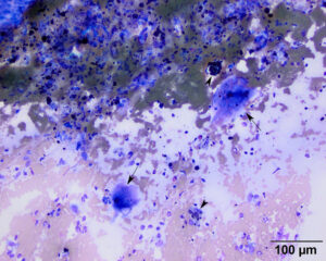
The aspirate was cellular and contained a mixture of inflammatory cells, many of which ere caught up within clots. At this low magnification, multinucleated giant cells, with hemosiderin in their cytoplasm (arrows), stand out, as do hemosiderin-containing macrophages (arrowheads) (20x objective, modified Wright’s stain).

