
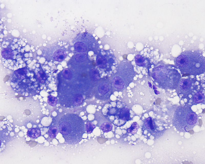
A direct smear of the liver aspirate revealed numerous hepatocytes with moderate to many variably sized but mostly small discrete margined cytoplasmic vacuoles, compatible with lipid accumulation (hepatic lipidosis). The nucleoli of the hepatocytes are also quite large and prominent, likely reflecting “reactivity” (modified Wright’s stain, 50x objective).