
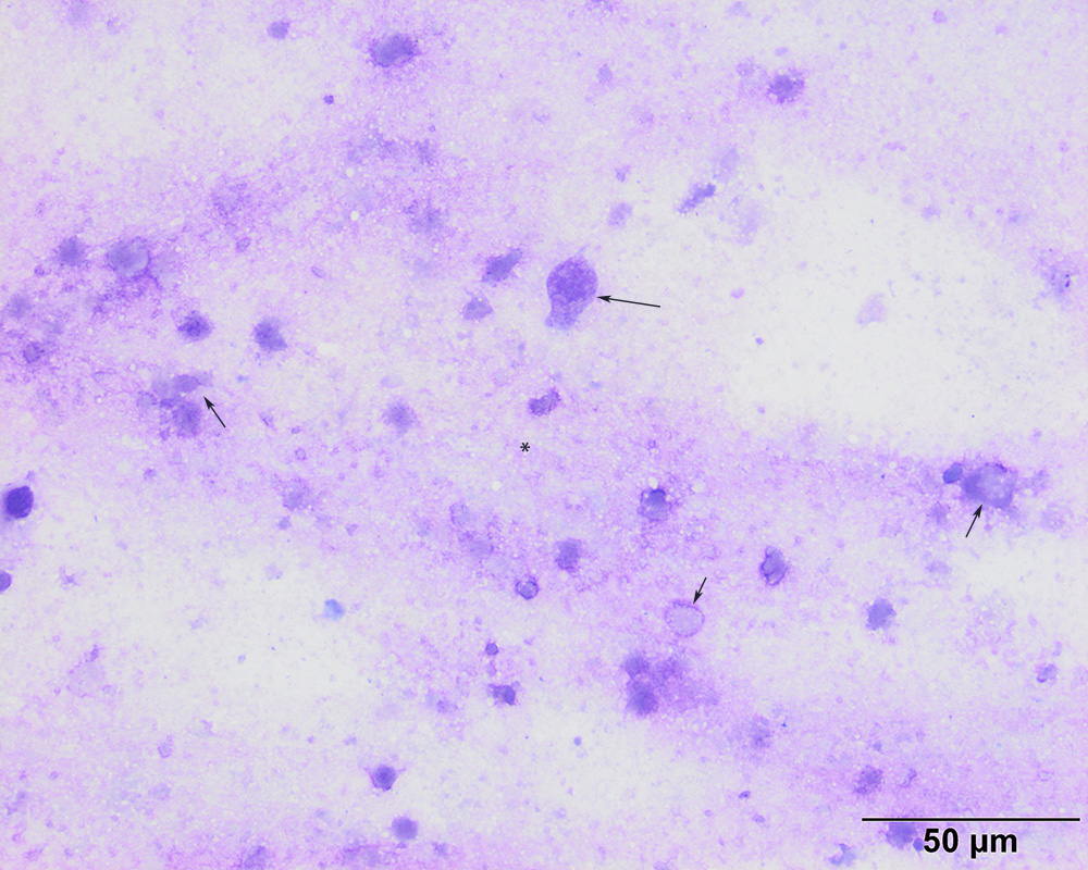
Numerous fragments from presumed dying cells are seen in the background, some of which have a round to cuboidal to columnar appearance (short arrows). The central cell has a distinct columnar appearance, with a fading nucleus (long arrow), supporting necrosis versus cell degeneration (modified-Wright’s stain, 100x objective)