
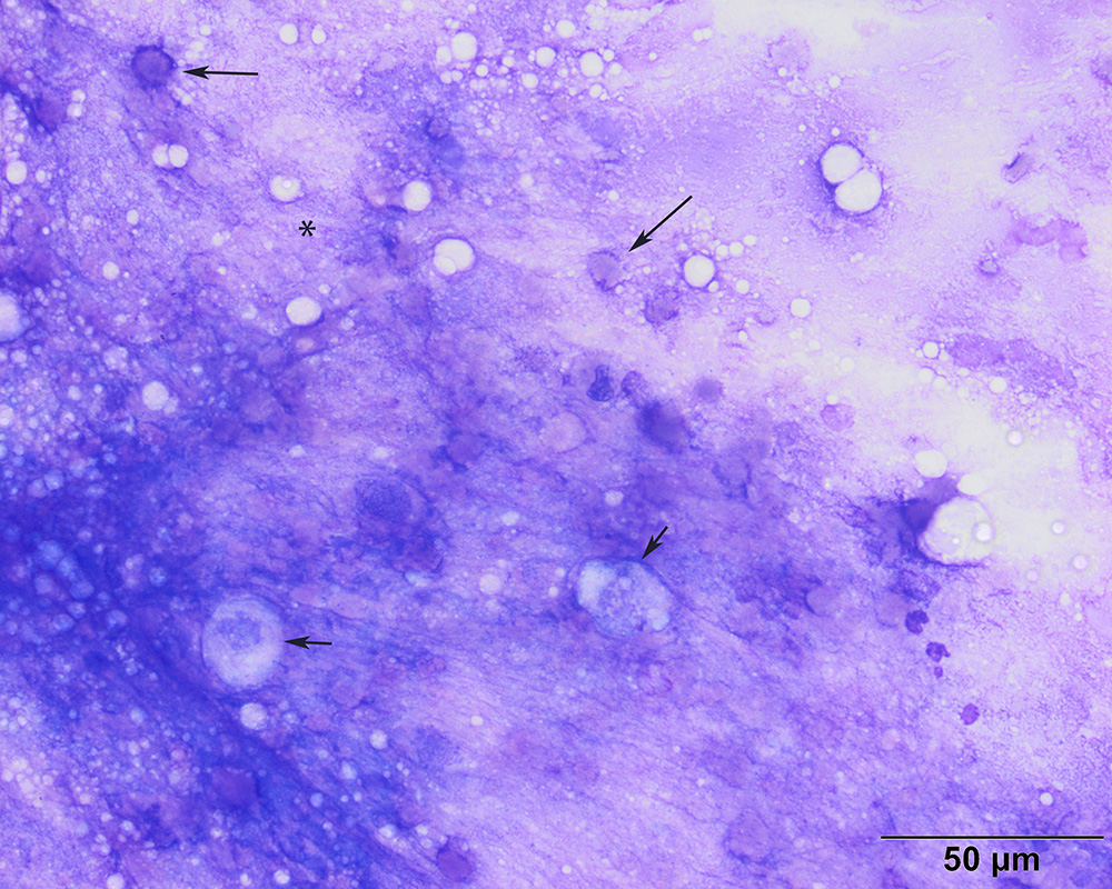
The mucus is thick and stringy (*) and the round structures resemble fragments of epithelial cells and were relatively uniform in size (arrows). Note the intact viable epithelial cells (short arrows). A disrupted neutrophil is also present in this image (modified-Wright’s stain, 50x objective).