
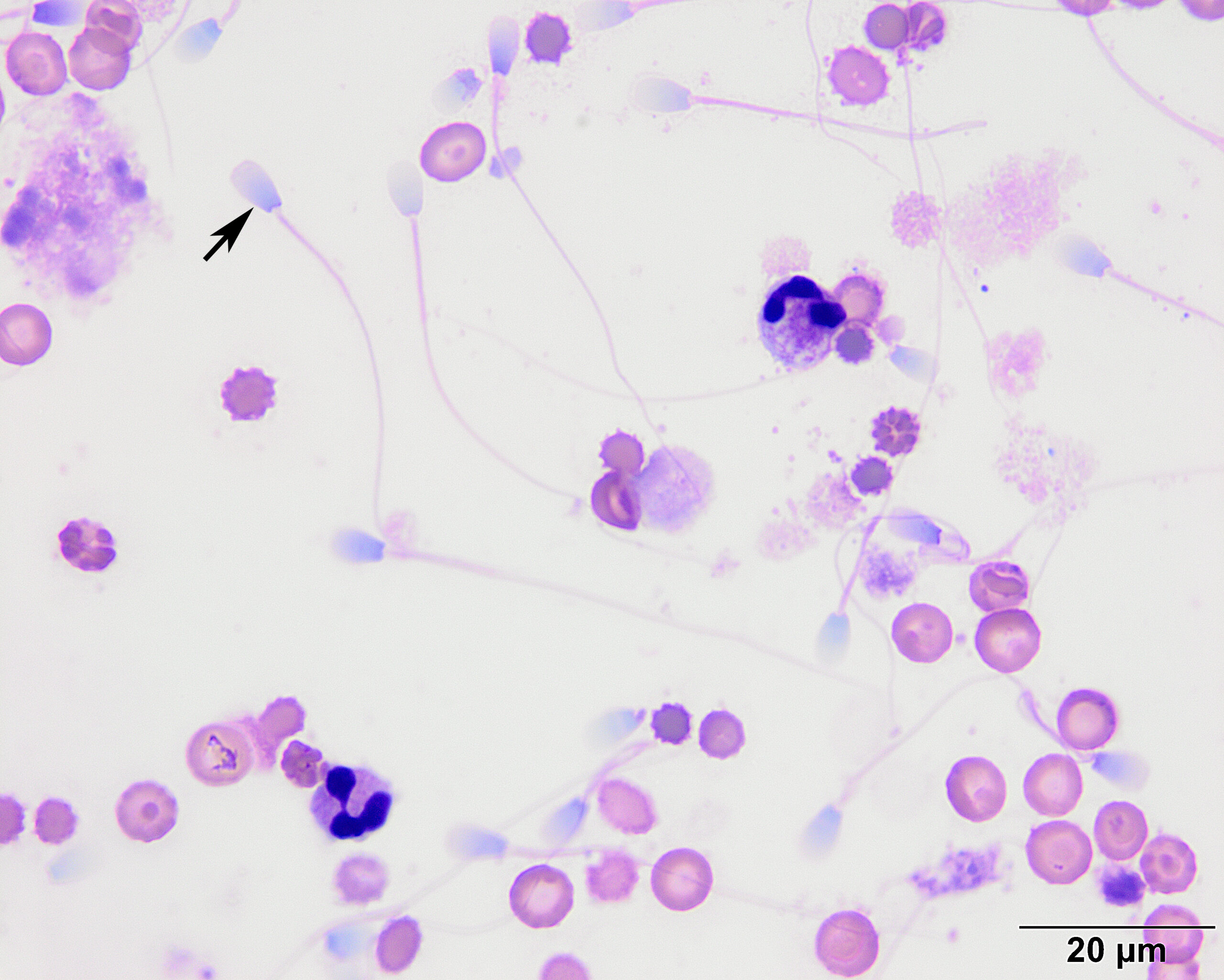
Higher magnification of cytospin smears showing spermatozoa (arrow) admixed with a few non-degenerate to raggedy appearing neutrophils and intact and lysing erythrocytes. There is a small amount of necrotic cellular debris, likely from trauma-induced injury (100x objective, Wright’s stain).