
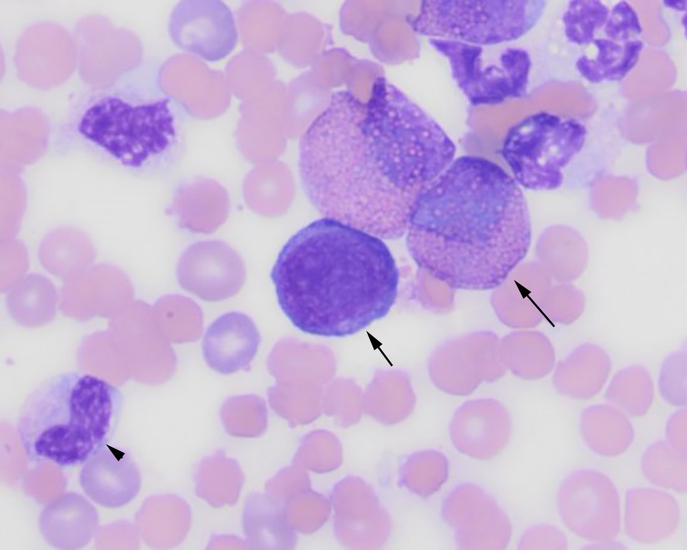
An eosinophilic myelocyte (long arrow), putative myeloblast (short arrow) and an immature neutrophil (metamyelocyte, arrowhead). The latter cell shows mild toxicity (Dohle body and cytoplasmic basophilia) (Wright’s stain, 50x objective).
An eosinophilic myelocyte (long arrow), putative myeloblast (short arrow) and an immature neutrophil (metamyelocyte, arrowhead). The latter cell shows mild toxicity (Dohle body and cytoplasmic basophilia) (Wright’s stain, 50x objective).
eClinpath helped 1.2 million visitors last year from 220 countries find important information on animal health. If you enjoy the site, please support our mission and consider a small gift to help us keep pace with its rapid growth. You can donate securely via PayPal or credit card. Thank you!
![]()