
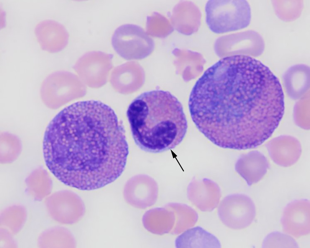
A band eosinophil (arrow) flanked by two eosinophilic myelocytes. Note the blue cytoplasm in the eosinophils and variable intensity of staining of the granules (Wright’s stain, 100x objective).
A band eosinophil (arrow) flanked by two eosinophilic myelocytes. Note the blue cytoplasm in the eosinophils and variable intensity of staining of the granules (Wright’s stain, 100x objective).
eClinpath helped 1.2 million visitors last year from 220 countries find important information on animal health. If you enjoy the site, please support our mission and consider a small gift to help us keep pace with its rapid growth. You can donate securely via PayPal or credit card. Thank you!
![]()