
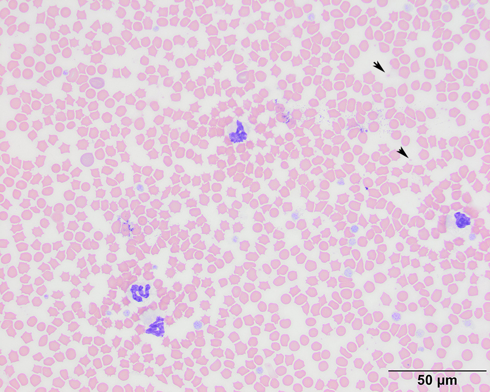
This low power image illustrates storage-associated changes in the leukocytes (neutrophils in this image), with swelling of the nuclear chromatin, forming band-like neutrophils (“pseudo” left shift). Echinocytes also likely reflect ATP depletion in red blood cells with storage. “Free” Heinz bodies are visible from this magnification in the background (arrowheads) (Wright’s stain, 20x objective).