
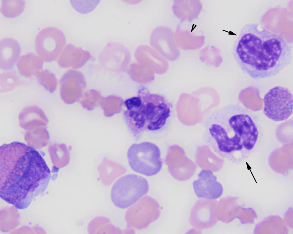
Eosinophils dominated in the smear and were mostly immature (myelocytes) with low numbers of mature segmented eosinophils. There was a left shift in neutrophils as seen by the band neutrophil in this image (long arrow), although mature neutrophils were the dominant cell in this lineage. There were also low numbers of monocytes (short arrow). Polychromatophils, acanthocytes and rare keratocytes (arrowhead) were evident in the erythrocytes (Wright’s stain, 50x objective).