
Aspirate of an interdigital swelling in a dog
Case Information
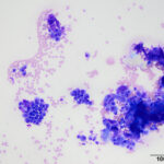
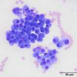
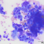
A 9 year-old, neutered male American pit bull terrier presented to a private veterinary practice for re-evaluation of a swelling within the interdigital space of P3 and P4 on the left front paw. The swelling was noted 6 weeks before, at which time it was oozing a hemorrhagic, purulent material. The swelling was initially thought to be an interdigital cyst and was treated with an course of antibiotics and a tapering dose of prednisone. The swelling seemed to shrink with treatment but did not resolve completely. At the re-evaluation, the veterinarian performed a fine needle aspirate of the swelling and submitted the smears of the aspirate to the Animal Health Diagnostic Center at Cornell University for examination. Examine the image from the smears below, then answer these questions:
- What broad category of tissue cells are present in the sample?
- Based on your answer for question 1, what would be your top differential diagnosis for the cause of the mass?
- What additional finding is evident in the smears that supports your answer to question 2?
|
|
|
|
Answers on next page