
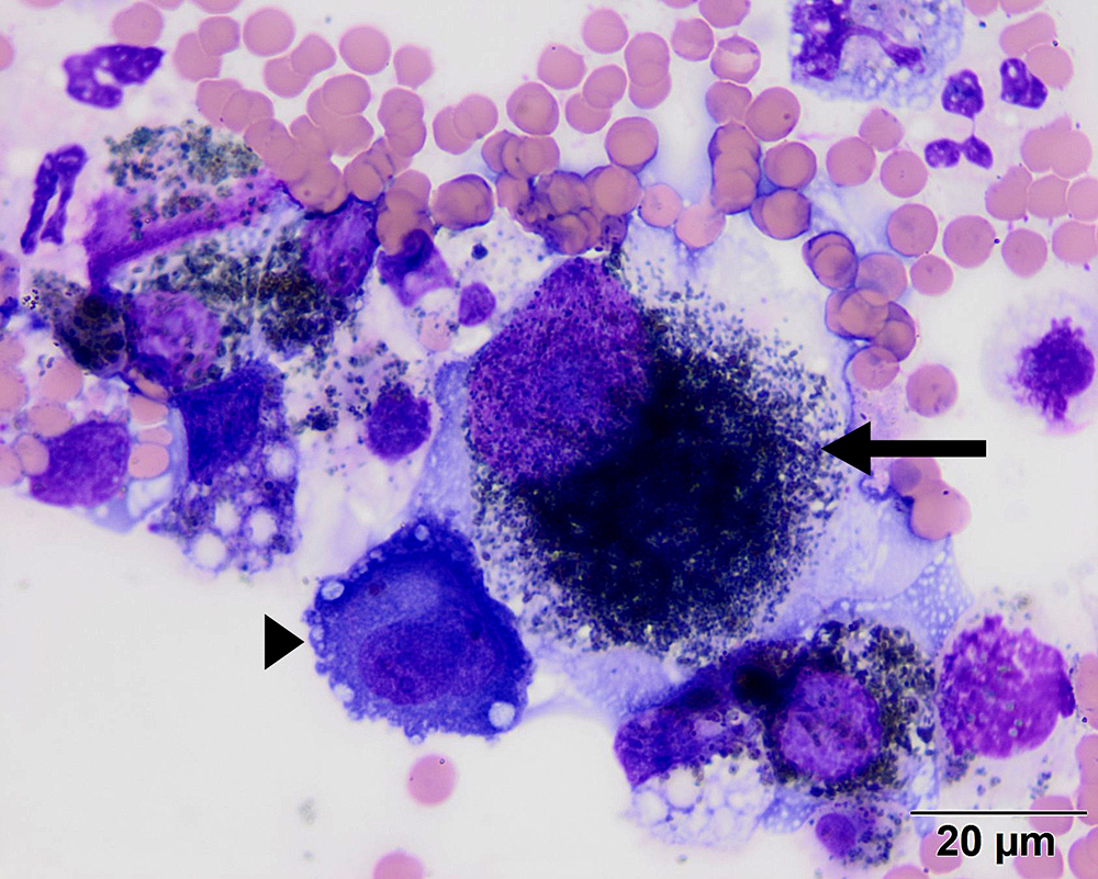
There are several individualized cells with eccentric round to oval nuclei and abundant melanin pigment, which were interpreted to be malignant melanocytes, with one very large neoplastic cell in the center (arrow). There are background inflammatory cells, including neutrophils, an erythrophagocytic macrophage and a plasma cell (arrowhead). (Wright’s stain, 100x objective)