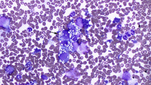
A linear cluster of flattened tumor cells have eccentric nuclei and abundant clear discrete-margined vacuoles (arrow); cytologic features that matched the tumor cells seen in the pleural fluid, supporting a malignant metastatic neoplasm. There were also increased neutrophils in the background with erythrophagocytic macrophages and necrosis (the latter two features not shown) (modified Wright’s stain, 50x objective).

