
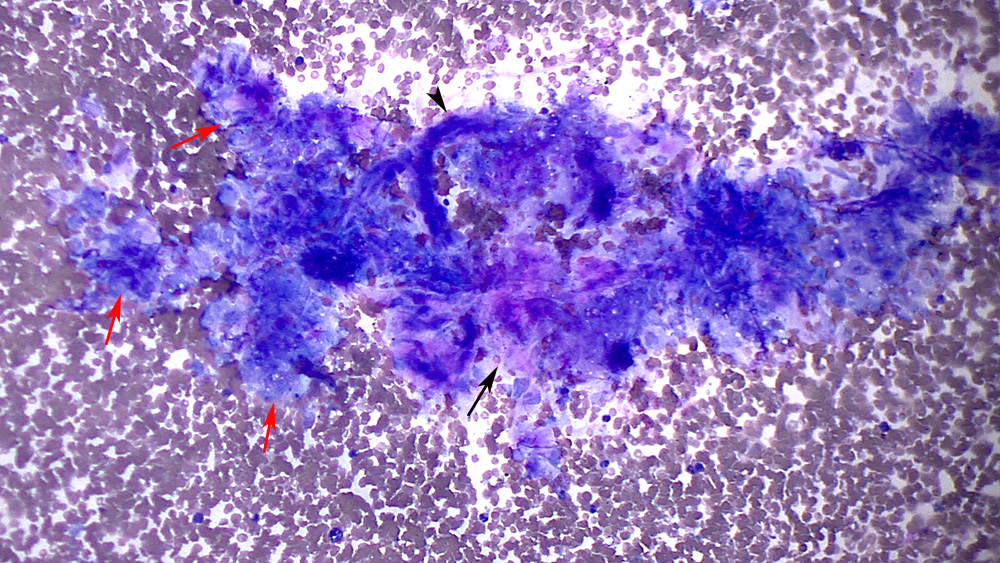
The aspirate contained numerous circular to linear clusters of medium to large tumor cells (red arrows) closely affiliated with aggregates of spindle cells (black arrow) and vascular profiles (black arrowhead). The spindle cells were caught up within a light pink smooth extracellular matrix. The latter could be a schirrous response, because the spindle cells were bland in appearance or a the stromal component of a granulosa cell tumor (suspected) (Wright’s stain, 20x objective).