
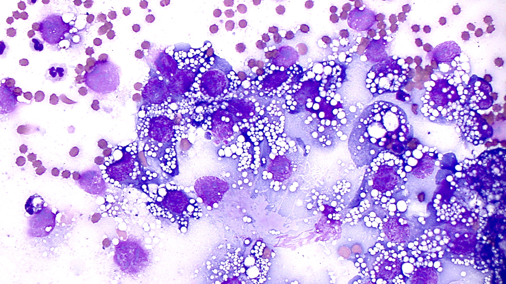
A sheet of vacuolated tumor cells was identified in a central streak within one of the sediment smears (modified Wright’s stain, 50x objective). Note the coarse chromatin and large irregularly shaped nucleoli in some of the cells.
A sheet of vacuolated tumor cells was identified in a central streak within one of the sediment smears (modified Wright’s stain, 50x objective). Note the coarse chromatin and large irregularly shaped nucleoli in some of the cells.
eClinpath helped 1.2 million visitors last year from 220 countries find important information on animal health. If you enjoy the site, please support our mission and consider a small gift to help us keep pace with its rapid growth. You can donate securely via PayPal or credit card. Thank you!
![]()