
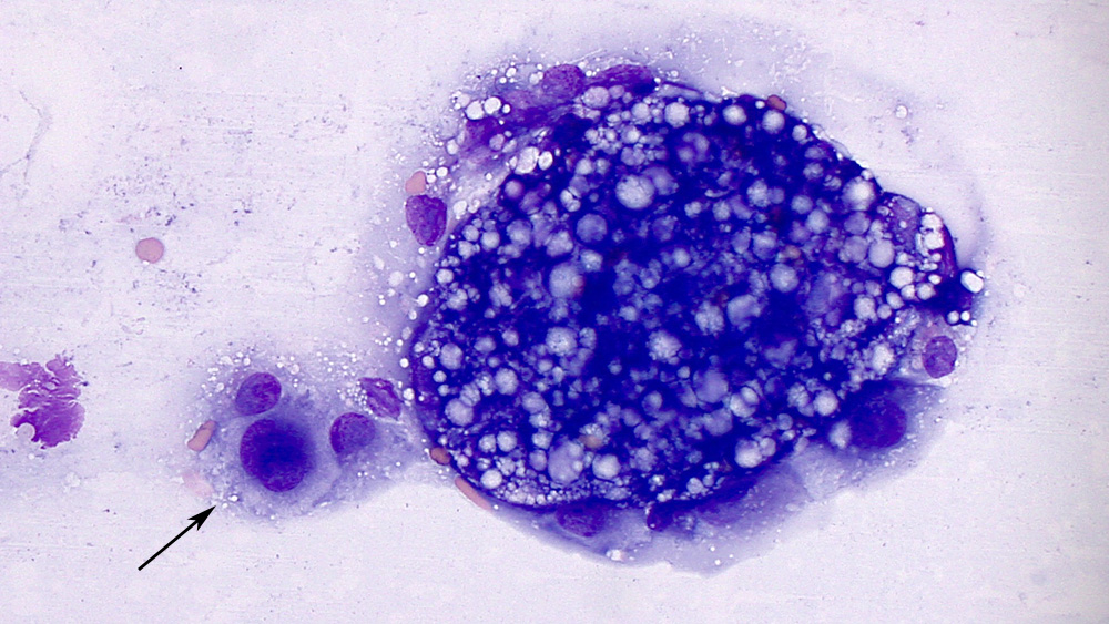
An individual mesothelial cell with a central round nucleus containing smooth chromatin (arrow) next to a cluster of vacuolated deep blue tumor cells at the feathered edge of the sediment smear (modified Wright’s stain, 50x objective).
An individual mesothelial cell with a central round nucleus containing smooth chromatin (arrow) next to a cluster of vacuolated deep blue tumor cells at the feathered edge of the sediment smear (modified Wright’s stain, 50x objective).
eClinpath helped 1.2 million visitors last year from 220 countries find important information on animal health. If you enjoy the site, please support our mission and consider a small gift to help us keep pace with its rapid growth. You can donate securely via PayPal or credit card. Thank you!
![]()