
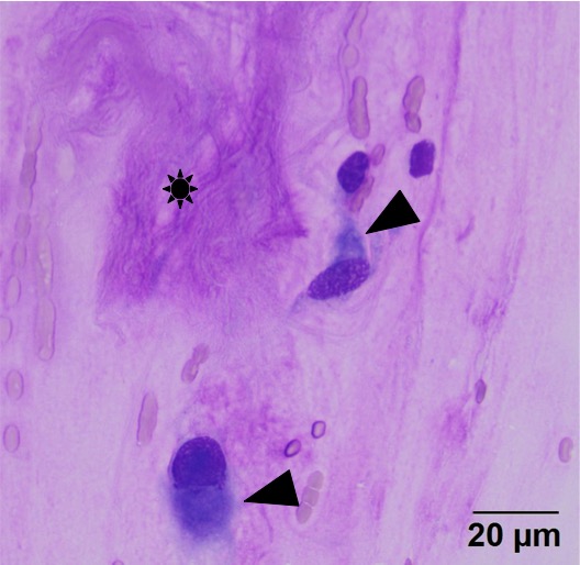
The spindle cells (arrowheads) display moderate anisocytosis and anisokaryosis with nuclear shapes ranging from round (lower cell) to ovoid (upper cell). They had moderate amounts of medium blue cytoplasm, that generally tapered away from both poles of the nuclei. Note the pink-purple larger fibrillar chunks of matrix material (mucin, presumptive, *) (Wright’s stain, 50x objective).