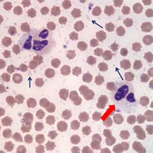
In this image taken of the blood smear prepared in the laboratory from stored blood (this was a mailed in sample), numerous small pink aggregates of coccobacilli, compatible with Mycplasma wenyonii, are observed in the background (black arrows). They can readily be mistaken for debris, but have a uniform structure. Some individual bacteria have classic ring shapes. Two neutrophils are also seen in this image (one labeled by the red arrow). Both cells are showing changes attributed to storage of blood in EDTA – polarization of pink neutrophil granules, a few discrete cytoplasmic vacuoles and nuclear swelling (contrast to cells in Figure 3) Red blood cells are also showing storage-associated changes, i.e. they are echinocytic (Wright’s stain, 1000x magnification).

